


Novel protocol to rapidly diagnose and treat retinal artery occlusion
Central retinal artery occlusion is characterized by painless, sudden vision loss in one eye, which may be a sign of an eye stroke.


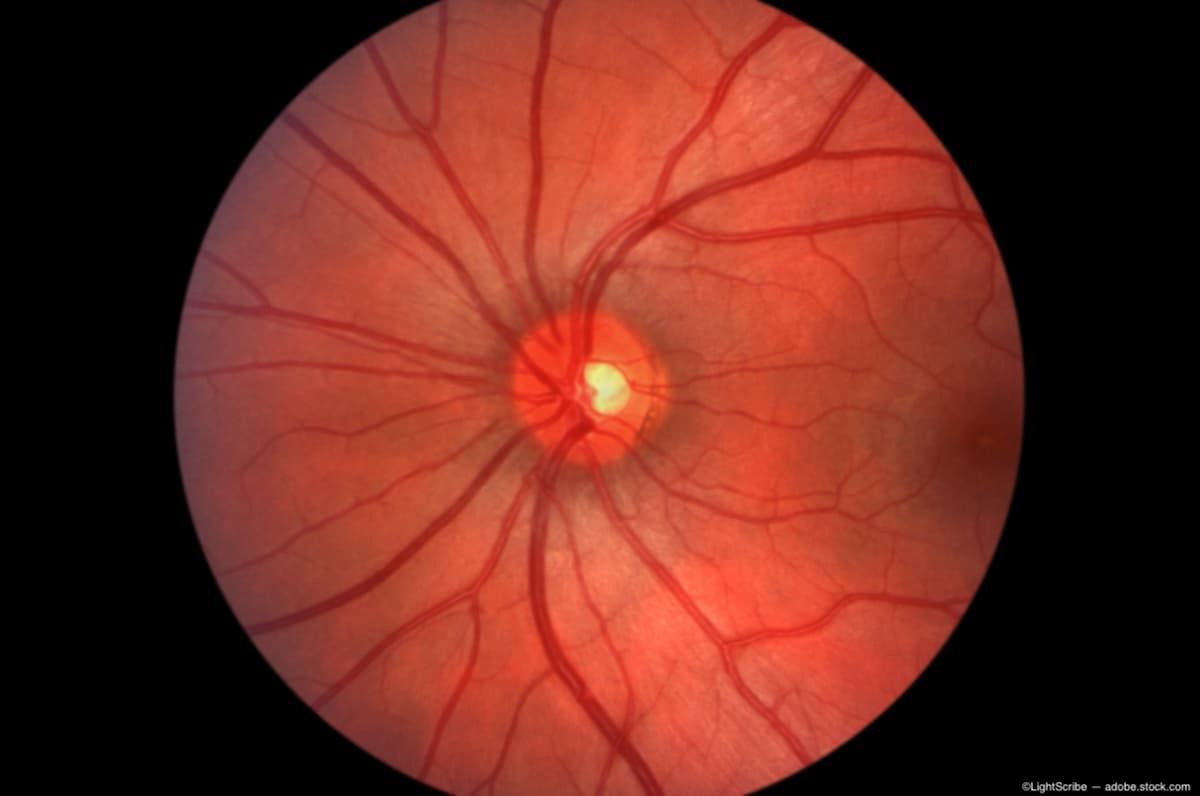
(Image credit: AdobeStock/LightScribe)
Ophthalmologists at New York Eye and Ear Infirmary (NYEE) of Mount Sinai, New York, have created a novel protocol to rapidly diagnose ocular strokes and provide emergency care to prevent irreversible vision loss. The authors, led by Gareth Lema, MD, reported their protocol in Ophthalmology.
Central retinal artery occlusion is characterized by painless, sudden vision loss in one eye, which may be a sign of an eye stroke. The vision loss occurs when the main artery to the retina becomes blocked, typically by a blood clot.
The urgency of this scenario is clear because the retinal nerve cells, which facilitate vision, die within hours following the occlusion unless the blood flow is reestablished. Ideally, the clot should be dissolved within 6 to 12 hours of the onset of vision loss, or the loss will become permanent.
The NYEE team uses high-resolution, semi-automated optical coherence tomography (OCT) retinal imaging in the emergency room in conjunction with rapid remote consultation to confirm the stroke diagnosis and expedite care, improving outcomes for patients with retinal artery occlusion and preserving vision.
The authors explained that by initiating care in the emergency department, the need for on-site ophthalmic consultants is reduced because those personnel may not always be readily available.
According to Richard B. Rosen, MD, Chief of the Retina Service for the Mount Sinai Health System, “Seamless cooperation and coordination between the subspecialty teams of stroke neurologists, retinal specialists, and interventional radiologists is the key to rapid triage of these emergency patients. This model can easily be implemented anywhere in the country where a stroke team is available.”
The protocol
The Department of Ophthalmology at Mt. Sinai launched an Eye Stroke Service and developed a treatment protocol for patients.
High-tech eye imaging devices using OCT were placed in 3 hospitals within the Mount Sinai Health System that have large emergency rooms and stroke teams.
When a patient with a possible retinal artery occlusion presents to the emergency room, the stroke service is urgently called to evaluate the patient and obtain OCT scans. The images are then sent electronically to remote on-call retinal specialists who can make an instant diagnosis. If the diagnosis of retinal artery occlusion is made, tissue plasminogen activator (tPA) is administered to the blocked ophthalmic artery to dissolve the clot.
The study included 59 patients who went through the protocol on the Eye Stroke Service during the first 18 months since it was launched. The investigators reported that the average time to treatment following arrival at the hospital was roughly 2.5 hours. Patients were treated an average of 9 hours following symptom onset; this time included the travel time to the hospital.
Of the 59 patients, 25 (42%) had a confirmed eye stroke based on OCT imaging and follow-up examination. Ten patients were eligible for treatment and 9 received treatment with intra-arterial tPA.
The investigators reported “a statistically significant improvement in the visual acuity 4 weeks after treatment. Researchers noted a dramatic improvement in 66% of these patients within 24 hours of treatment, with 56% of treated patients maintaining improved vision 1 month after the procedure. The patients’ vision improved dramatically from count fingers to 20/100. “Most notably, four patients, 44% of those treated with tPA, improved to 20/40 or better, which constitutes unrestricted driving vision in New York State,” the investigators commented.
The goal going forward is to improve the outcomes by expanding the availability of OCT cameras in more Mount Sinai Health System emergency rooms and increasing outreach to the medical community and general public. According to the press release, another goal is for people to better understand both the symptoms of eye stroke and the importance of getting immediate medical attention, and recognize the potential for treatment to preserve vision with a timely referral.”
Lema emphasized, “The importance of education among the medical community and the public cannot be stressed enough. Patients with an eye stroke are at risk of also developing a stroke in the brain, and that risk is highest in the first month after diagnosis. If patients get to us in time, we can treat. But even for those who don’t, the systemic work up to identify the source of the stroke might be even more critical.”