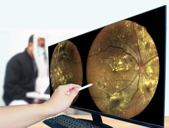
Diabetic eye disease: Best treatment options for your patients
Although a number of medical treatment advances for diabetic retinopathy (DR) and diabetic macular edema (DME) have occurred in recent years-predominantly through the introduction of the anti-vascular endothelial growth factor (VEGF) agents-controlling the systemic underlying disease is of paramount importance.
Even patients with good-quality vision who are asymptomatic must understand DR/DME could become symptomatic quickly.
Therefore, the American Academy of Ophthalmology (AAO) recommends diabetic patients have annual comprehensive dilated eye exams at minimum.1
AAO endorses a step-wise approach to the management of diabetic retinopathy and DME depending on disease stage. Patients should be followed closely with routine exams and imaging, such as optical coherence tomography (OCT), fluorescein angiography, and optical coherence tomography angiography (OCTA), to assess disease severity.
Treatments for DR and DME include laser treatment (focal, grid, and panretinal), intravitreal anti-VEGF therapy, and steroids.
Laser treatments
Laser photocoagulation has been the standard of care for DME for more than 30 years, based on the ETDRS protocol.2,3
Focal laser is used to treat noncenter-involved DME in areas of leaking microaneurysms between 500 μm and 3,000 μm from the center of the macula.
Grid laser, on the other hand, is used to treat diffuse DME and can be placed more than 500 μm from the center of the macula and more than 500 μm from the temporal margin. Complications from both include loss of central vision from scaring.
Panretinal photocoagulation is recommended for patients with DME, with or without neovascularization, and severe peripheral ischemia at level 53 or worse, according to the ETDRS severity scale.
During the procedure, thermal burns are created in the peripheral retina, resulting in tissue coagulation and improved retinal oxygenation. Complications include abnormal fluid accumulation, retinal detachment, and visual field and night vision defects.
Laser therapy slows disease progression and vision loss, but it does not improve vision. Although laser therapy is still used in the clinical management of these diseases, the development and approval of anti-VEGF agents have completely revolutionized the treatment of diabetic retinopathy and DME.
Anti-VEGF therapy
Currently, anti-VEGF therapy is the recommended treatment for DR and center-involved DME. To date, the FDA has approved two agents for this indication: ranibizumab, and aflibercept; a third (bevacizumab) is often used off-label.4-6
The AAO recommends that all patients who receive anti-VEGF injections be examined 1-month post-therapy.1 Complications from anti-VEGF therapy are rare, but can be severe, and include infectious endophthalmitis, cataracts, and retinal detachment.
The biggest challenge surrounding anti-VEGF treatment is the treatment burden on both physicians and patients. Clinical trials have shown that anti-VEGFs work best when given consistently every few weeks, which can be inconvenient to patients in terms of time, money, and transportation to and from appointments, especially because many patients don’t experience visual improvement. These factors frequently lead to noncompliance and disease progression.
As such, a need for longer-acting anti-VEGF agents have been identified, and clinical trials are currently under way.
Steroids
When laser treatment and anti-VEGF injections fail, many retinal specialists turn to intravitreal corticosteroids as an additive for the treatment of persistent DME.
Patients who have had less than a 50% reduction in macular thickness from baseline after 3 or 4 monthly anti-VEGF injections are candidates for steroids, as well as patients whose visual acuity has not improved to 20/40 after 3 to 6 monthly anti-VEGF injections because of edema.7
Available agents include triamcinolone acetonide, dexamethasone, and fluocinolone acetonide, but only dexamethasone and fluocinolone acetonide have been approved by the FDA for this indication. Steroid treatment is not without risks, and include cataracts and glaucoma. Glaucoma development is of particular concern; therefore, patients undergoing steroid therapy should have their IOP closely monitored.
Takeaways
Although the treatment of DR and DME has never been more robust, clinicians agree that prevention through proper diabetes management is key. Once retinal disease does develop, early intervention is critical to prevent further vision loss.
References:
1. American Academy of Ophthalmology Retina Panel. Preferred Practice Pattern Guidelines: Diabetic Retinopathy. San Francisco, CA 2016.
2. Photocoagulation for diabetic macular edema. Early Treatment Diabetic Retinopathy Study report number 1. Early Treatment Diabetic Retinopathy Study research group. Arch Ophthalmol. 1985;103:1796-1806.
3. Relhan N, Flynn HW, Jr. The Early Treatment Diabetic Retinopathy Study historical review and relevance to today's management of diabetic macular edema. Curr Opin Ophthalmol. 2017;28:205-212.
4. Nguyen QD, Brown DM, Marcus DM, et al. Ranibizumab for diabetic macular edema: results from 2 phase III randomized trials: RISE and RIDE. Ophthalmology. 2012;119:789-801.
5. Michaelides M, Kaines A, Hamilton RD, et al. A prospective randomized trial of intravitreal bevacizumab or laser therapy in the management of diabetic macular edema (BOLT study) 12-month data: report 2. Ophthalmology. 2010;117:1078-86 e2.
6. Korobelnik JF, Do DV, Schmidt-Erfurth U, et al. Intravitreal Aflibercept for Diabetic Macular Edema. Ophthalmology. 2014;21:2247-2254.
7. Regillo CD, Callanan DG, Do DV, et al. Use of Corticosteroids in the Treatment of Patients With Diabetic Macular Edema Who Have a Suboptimal Response to Anti-VEGF: Recommendations of an Expert Panel. Ophthalmic Surg Lasers Imaging Retina. 2017;48:291-301.
Newsletter
Keep your retina practice on the forefront—subscribe for expert analysis and emerging trends in retinal disease management.


