
News|Articles|September 20, 2024
EURETINA 2024: Ricardo Leitão Guerra, MD, MSc, FICO, speaks about multimodal imaging
Author(s)Hattie Hayes, Martin David Harp
Why having a set process for diagnostic imaging is crucial to patient outcomes
Advertisement
Ahead of this year’s European Society of Retina Specialists (EURETINA) meeting, we sat down with Ricardo Leitão Guerra, MD, MSc, FICO, to discuss a presentation he’ll be giving as part of the industry satellite symposium on Friday, 20 September. Dr Guerra is from Clínica de Olhos Leitão Guerra in Salvador, Bahia, Brazil.
He talked about developing a set process for retinal imaging, how he fits together the “puzzle pieces” of OCT and other modalities, and introducing an artificial intelligence (AI) component from ZEISS into his clinical workflow. Watch the full interview for his approach to workflow.
Editor's note: The below transcript has been lightly edited for clarity.
Hattie Hayes: Hi. My name is Hattie Hayes, and I'm the editor of Ophthalmology Times Europe. This year, the European Society of Retina Specialists is holding its congress in Barcelona, Spain. Joining me today to talk about one of his presentations, we have Dr Ricardo Guerra. Thank you so much for being here with me. I'm really excited to speak with you.
Ricardo Leitão Guerra, MD, MSc, FICO: Thank you for the invitation. It's great to talk a little bit about the presentation.
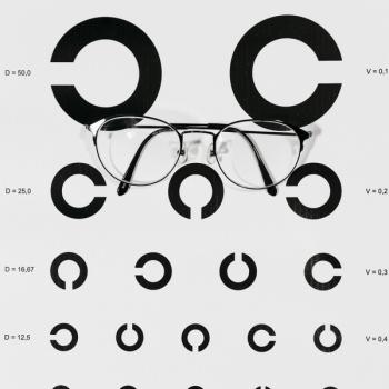
HH: Wonderful. Now, I know that you're speaking about multimodal imaging at the Industry Satellite Symposium. Tell me a little bit about the landscape of retinal imaging in 2024.
RG: So, I think that we have, now, access to so many kinds of imaging modalities that we kind of lose information from the basics. I don't think that we have a better imaging modality if it…[doesn’t] come from research, but every imaging modality is kind of a piece of a puzzle.
I think that understanding all of them, the power of each one of these modalities, is kind of the landscape nowadays.
HH: I like that description of it as pieces of a puzzle. Could you talk a little bit more just about that analogy and how that factors into your own clinical practice?
RG: In my daily practice, I use ZEISS devices, as I will talk about in the symposium. I use a software to analyse each patient for every kind of imaging modality. So I have fundus photography, to understand the status of the eye, and then I can split the monochromatic channels and get more information for specific layers. Then I can understand if the patient needs a fluorescein angiography or an OCT.
And then, understanding the patient, understanding the status of the retina, I can go forward to the next piece of the puzzle. In the end what we want is to get the most information possible to get to the correct diagnosis and take care of that patient in the best way.
HH: That's good. And it is such a comprehensive, multistep process. I'm glad that you are talking about your own clinical practice. What do you think your fellow clinicians misunderstand or fail to consider in their own approach to diagnostic imaging?
RG: Well, I used to receive patients from other colleagues, good ones. And sometimes they failed to identify the disease because they didn’t have access to the device that the imaging model [required]. For example, in OCT, you get the reports. However, [a particular] patient went to up to 128 B-scans. We have to analyse each one of these B-scans to get the comprehensive care of that patient. And most clinicians don't have the opportunity to to work with softwares and devices, and…this is the main problem, the main cause of good physicians failing to to identify or to diagnose a disease.
HH: Having the right tools is so important, and it's something that you owe to your patients. Yeah. And I I like that phrasing, too, of “good physicians failing to diagnose the disease.
Now, I'm curious to know one thing I like to ask clinicians is what they consider to be kind of the keyword or the catchphrase of their industry. So when we're talking about retinal care, if you had to sum up the retinal care industry in one word or phrase, what would you pick and why?
RG: I think that the key[word] is…”process,” kind of a best-practice process. Always do the same steps. Don't skip steps. Even when you think, “This patient doesn't need this step,” you have to always follow the steps…When we have our best practices and we always do things the same way, we have less chance of failing, or not identifying the disease. Taking care of our patients is kind of our responsibility. And there is no space...to fail in diagnosing.
HH: I think that's great when, you know, when you have a specific process or routine in your practice, then you get to customise the care to the patient, and that leaves out so much less room for error.
RG: Yes. By the way, I have a practice for analysing the OCT. Every OCT, I follow the same steps: I look at the B-scans, the main B-scans, high quality, then I look at the maps for each segmentation, ellipsoid zone, mid retina, vitreous interface.
Then I go to analysis of the B-scans, the 128 B-scans. It makes my process, or my best practice of identification, faster and takes less effort. And it's kind of a way that I teach all my fellows and residents that are with us. I had access recently to a new device, a new software from ZEISS. It's called PathFinder, and what this software does is uses AI. It identifies the B-scans that I have to look at. And I will have to remake my best practice process in analysing OCT! But I think that, now, it will be faster.
HH: Well, “process” and “progress” are only just a couple letters away! It's so important to be adaptable even when you have that routine. Right?
Was there anything that you're excited to talk about on [Friday] that we haven't touched on here today?
RG: Well, I'm kind of very excited about some new information that we found, regarding blue light reflectance. It's kind of an old kind of imaging modality, costs less and is available in every kind of device that can form fundus photography.
And we researched some good information, some new information, and we are currently under review for publishing it. We now can identify…midretina abnormalities using a color fundus photography composite in the blue channel.
And, we are writing about it, and we are excited. I will show, at this symposium, some of our results, and I think it will help so many physicians and patients all around the globe. It's inexpensive. Everybody can do it and it can help others in their daily practice.
HH: Well, you've really given some good, I think, pearls for anyone watching to take home and take back to their practice post conference. So thank you so much for speaking with me and for speaking to our audience today. Have a fun, safe trip to Barcelona.
RG: Thank you.
Newsletter
Keep your retina practice on the forefront—subscribe for expert analysis and emerging trends in retinal disease management.
Advertisement
Related Content
Advertisement


Latest CME


Advertisement
Advertisement