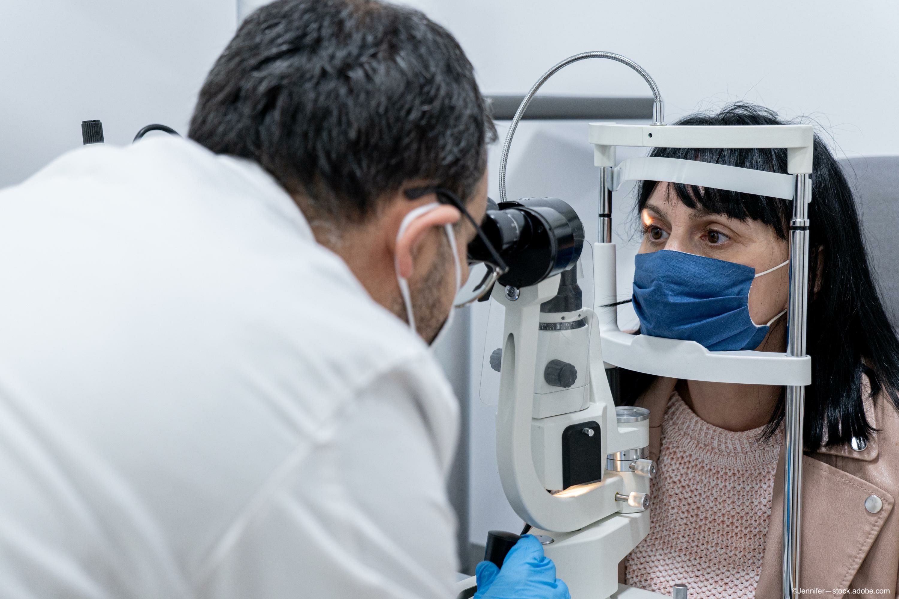
First signs of COVID-19 seen in the retina, investigators report
A team of investigators found that the retina may offer signs of COVID-19 infection before symptoms present.

According to investigators from Singapore National Eye Centre in Singapore, the retina may show signs of COVID-19 infection even before symptoms appear in the rest of the body.
The researchers theorized that “the retinal microvascular signs may result from cardiovascular and thrombotic alterations associated with COVID-19 infection.”
Related: The impact of COVID-19 on the management of wet AMD
The team conducted a prospective cross-sectional study,1 which included patients who tested positive for COVID-19 by nasopharyngeal swab and had no medical history. The patients then underwent retinal imaging using optical coherence tomography (OCT) that provided color fundus photographs (CFP) and macular scans, according to Ian Yeo, MBBS, FRCS, corresponding author,
A total of 108 patients (216 eyes; mean age, 36.0±5.4 years) were included. Of these, 41 (38.0%) patients had acute respiratory infection symptoms when they presented.
Related: Adapting medical retina service in COVID-19 era requires careful approach
The authors reported that 25 (11.6%) eyes had retinal signs of infection in the CFP and/or OCT images. Nine patients had bilateral retinal findings that included 8 eyes (3.7%) with microhemorrhages, 6 eyes (2.8%) with retinal vascular tortuosity, and 2 eyes (0.93%) with cotton wool spots. OCT showed hyperreflective plaques in 11 (5.1%) eyes in the ganglion cell layer, and 2 of these eyes also had retinal signs on the CFP (cotton wool spots and microhemorrhages, respectively).
Investigators did not observe significant (p = 0.227) differences in the prevalence rates of the retinal signs between symptomatic and asymptomatic patients. However, the patients with retinal signs were “significantly more likely to have transiently elevated blood pressure than those without (p = 0.03).”
Reference
1. Sim R, Cheung G, Ting D, et al. Retinal microvascular signs in COVID-19. Br J Ophthalmology 2021; http://dx.doi.org/10.1136/bjophthalmol-2020-318236
Related Content: Diabetic Macular Edema | AMD | Diabetic Retinopathy
Newsletter
Keep your retina practice on the forefront—subscribe for expert analysis and emerging trends in retinal disease management.