Functional microglia derived from human pluripotent stem cells in retinal organoids
By co-culturing the differentiated microglia with retinal organoids, investigators found they may be able to recapitulate retinal development.
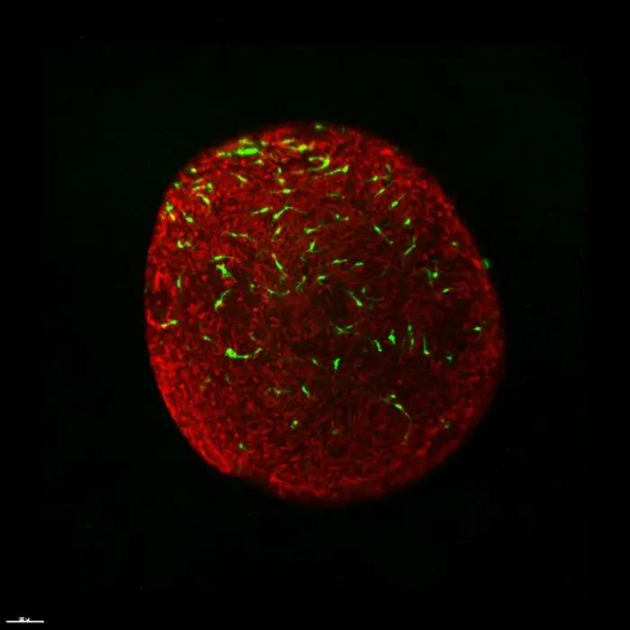
Reconstructed 3D images showed that microglia were located underneath the photoreceptor cell layer. Green - microglia; Red - photoreceptor cells. (Image courtesy of China Science Press)

Zi-Bing Jin, MD, PhD, of the Beijing Institute of Ophthalmology, led a team of researchers from the institute as well as Beijing Tongren Eye Center, Beijing Tongren Hospital and Capital Medical University in research on the retinal organoids, which could faithfully recapitulate retinal development.
“Retinal organoids provide an excellent research tool to address fundamental questions, understand disease delineation and discover new drugs.” Jin said in a news release.
However, microglia, the key neuro-immune cells regulating the natural microenvironment of the retina, are completely absent from the currently developed retinal organoids, which made the organoids less representative of real retinas.
According to the news release, the team found that there are published protocols for macrophage and brain microglia differentiation.
“We try to generate retinal microglia in our lab, because the macrophage and microglia are all from hematopoietic progenitors” he said in the news release. “Finally we get it, a very simple and high efficiency method to produce retinal microglia.”
The investigators also noticed that the differentiated microglia could distinguish deteriorated cells or cell debris from healthy cells and cleanup the surrounding microenvironment by phagocytosis. They are capable of the same function of in vivo microglia.
The researchers keep trying another amazing idea. They co-culture the differentiated microglia with retinal organoids.
“No one has ever done that before. It is really awesome when I see the microglia migrate to the right position, proliferate and become retina-resident microglia in 3D retinal organoids.” Jin explained in the news release.
These new exciting results prove that the team generates “smart” microglia, which could move into retinal organoids and consciously “find their own way around.” They are precisely positioned and functionally accurate. They make the retinal organoids closer to the retinal organ.
The team provided a more efficient protocol to generate microglia from human pluripotent cells and proof-of-concept evidence that these microglia could be further differentiate into retina-resident microglia in a 3-D retinal niche, presenting a new toolkit of “integral retinal organs” for studying retinal microglial biology, retinal development, and retinal regenerative diseases.