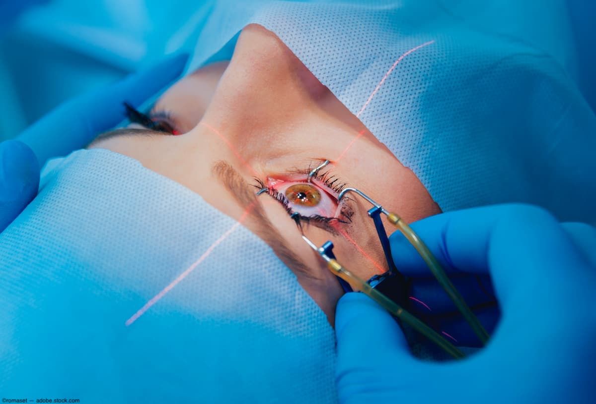
Laser retinopexy during primary scleral buckling
With the newly modified lighted endolaser probe, an 18-gauge angiocatheter sleeve protects against vitreous disturbance.
With the newly modified lighted endolaser probe, an 18-gauge angiocatheter sleeve protects against vitreous disturbance. (Image Credit: Adobe Stock/romaset)

A modified illuminated endolaser is helping to facilitate safe endophotocoagulation during chandelier-assisted scleral buckling surgery. Surgeons from the New York Eye and Ear Infirmary of Mount Sinai, New York, reported on a case series of patients in which the revamped instrument was used.
In this series, 6 phakic patients with rhegmatogenous retinal detachments (RRDs) underwent primary scleral buckling with chandelier endoillumination, external drainage, and endophotocoagulation using the modified endolaser.
The researchers, led by first author Nilesh Raval, MD, explained that retinopexy during scleral buckling is typically performed using indirect laser or cryotherapy and can be done under indirect visualization or using chandelier endoillumination.
With the newly modified lighted endolaser probe, an 18-gauge angiocatheter sleeve protects against vitreous disturbance during scleral buckle retinopexy under microscopy visualization.
Case report
A representative case was that of a 17-year-old highly myopic phakic female who presented with a fovea-splitting, inferotemporal RRD with multiple inferior breaks. The surgeons positioned a primary scleral buckle para-equatorially 1 buckle width posterior to the rectus muscle insertions to support the inferior breaks. A chandelier was inserted through a 25-gauge valved trocar 180 degrees from the area of pathology to illuminate the fundus and confirm the inferior break locations and subretinal fluid was drained.
The surgeons trimmed an 18-gauge angiocatheter to 20 mm long and slid it over the 27-mm lighted endolaser probe, leaving 7 mm of the endolaser probe exposed.
The authors commented, “Given that a 7-mm chandelier can be inserted safely through a valved trocar into a nonvitrectomized eye without causing vitreoretinal traction, the modified laser probe was conceived as an equivalent substitute for the chandelier tip. With the sheathed, lighted endolaser inserted into the superiorly placed trocar, laser retinopexy was applied around the inferior breaks at power settings between 400 and 500 mW.”
The procedure ended with injection of a 0.4-cc bubble of 100% sterile sulfur hexafluoride gas through the pars plana and sclerotomy and conjunctival closure with plain gut sutures.
The other 5 patients with phakic RRDs underwent similar chandelier scleral buckling procedures with guarded laser photocoagulation. All 6 procedures were successful.
The surgeons concluded that the new endolaser setup was safe and efficient for endophotocoagulation during chandelier scleral buckling. “This instrument is successful in creating adequate retinopexy without disturbing the vitreous or contacting the lens in young phakic patients. We recommend using this setup as an alternative to cryotherapy or indirect laser retinopexy during primary scleral buckling with chandelier-assisted endoillumination.”
Reference
1. Raval N, Li Y, Pinhas A, et al. Modified endolaser instrument for chandelier scleral buckling. J Vitreoret Surg. 2023; doi: 10.1177/24741264231171243journals.sagepub.com/home/jvrd
Newsletter
Keep your retina practice on the forefront—subscribe for expert analysis and emerging trends in retinal disease management.