Multicolor and autofluorescence imaging: coming to terms
Technologies offer different approaches to different scenarios
This article was reviewed by Giovanni Staurenghi, MD.
Imaging technology has advanced markedly and provides different approaches to visualizing different scenarios, according to Giovanni Staurenghi, MD, who described the practical applications of multimodal imaging.
Available modalities depend on white light from a standard flash nonconfocal camera, as in conventional fundus cameras (Canon and Topcon); a scanning laser with 2 wavelengths (Optos); slit-light confocal light-emitting diodes (LEDs; Eidon, CenterVue); broad line fundus images using blue, green, and red LEDs (Zeiss Clarus, Carl Zeiss Meditec); a scanning confocal laser with 3 wavelengths of blue, green, and near-infrared (IR; Spectralis, Heidelberg Engineering); and a scanning confocal laser with 3 wavelengths of blue, green, and red (Scanning Laser Ophthalmoscope Mirante, Nidek).
The main difference between the normal fundus and multicolor images is the use of near IR reflectance instead of red and the confocal aperture.
Staurenghi, a professor of ophthalmology in the Department of Biomedical and Clinical Science at Luigi Sacco Hospital, University of Milan in Italy, noted that once a normal fundus image is split into red-green-blue channels, the red image of the retinal pigment detachment shows high reflectance. Meanwhile, the near IR (NIR) reflectance component of a multicolor image of the same case shows a dark area because of the absorption of the IR light. Thus, when the red channel is produced by IR light, only blue and green light are visualized. The confocal aperture provides higher definition and sharp images.
Moreover, as the focuses of the NIR light and the blue and green differ, the image colors can change depending on the degree of focus. In 2 images obtained from the same patient on the same day with scan focuses of -2.37 D and -0.72 D, the details differed completely in sharpness and color.
Multicolor imaging
Results from a number of studies have reported on the importance of multicolor images in identifying specific features such as pseudodrusen and atrophy compared with other imaging modalities. The use of a confocal aperture is a key component in the superiority of multicolor images.
“The confocal aperture allows visualization of only the light from the area of interest and not that coming from the surrounding area,” Staurenghi said. “This capability results in a markedly increased sharpness of the images.”
He described 4 images of geographic atrophy, 2 of which were obtained with confocal instruments (Eidon, CenterVue; Spectralis, Heidelberg Engineering), 1 with a normal fundus camera (Kowa American Corp), and 1 with a blue auto-fluorescence image obtained with the Spectralis. The sharpness obtained using the confocal aperture enables the identification of choroidal neovascularization, which is not visible using nonconfocal color fundus images or autofluorescence.
The use of single-wavelength green, blue, or NIR light coupled with the confocal aperture allows better definition of single structures such as the nerve fiber layer (blue) and retinal pigment epithelium (green), and deeper structures such as the choroidal vasculature.
Autofluorescence Imaging
This technology, Staurenghi explained, uses the fluorescence emanating from lipofuscin in the retinal pigment epithelial cells. Numerous available instruments can produce autofluorescence images; however, he pointed out that blue light is obtained from the confocality of the instruments and green from other instruments.
“Confocality is important in this case because all the autofluorescence light is collected from the entire eye, including the lens, which can interfere with fundus visualization,” he said. “The use of confocality with the possibility to obtain only the light coming from the layer of interest permits visualization of the fundus autofluorescence. For the green autofluorescence, the absorption and fluorescence from the lens are less, and instruments without a confocal aperture also can obtain an autofluorescence fundus image.”
The blue light visualizes the macular pigment, which is yellow and absorbs blue light.
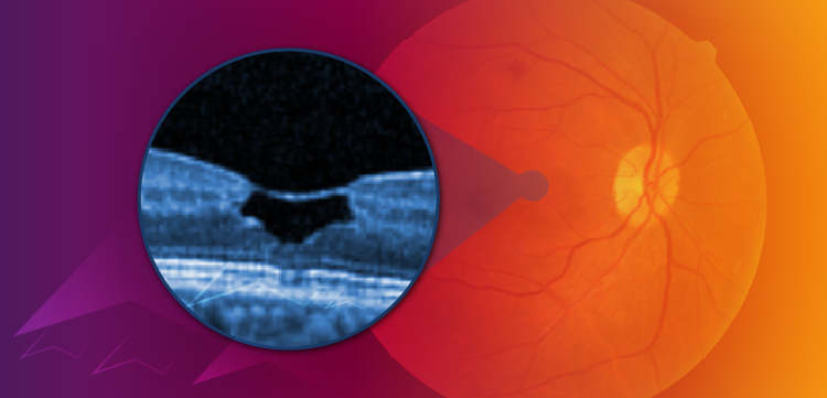
Staurenghi explained that blue autofluorescence can be used to identify macular atrophy, pseudoholes, lamellar holes, and macular holes before and after surgery. For multiple evanescent white dot syndrome, an inflammatory disease, autofluorescence imaging shows bleaching of the characteristic white dots after treatment. In inflammatory choroidal neo-vascularization, hyperautofluorescence is always apparent. In the expansion of serpiginous choroiditis, the enlarging area is always hyperautofluorescent before progressing to atrophy.
Conclusion
“Multicolor imaging provides the sharpest images due to the confocal aperture and better visualization of some pathologies because of the IR reflectance component,” he concluded. “Autofluorescence imaging is the best technology to detect the retinal pigment epithelium and is important for the differential diagnosis in macular atrophy, macular holes, pseudoholes, and lamellar hole and inflammatory diseases.”
Giovanni Staurenghi, MD
E: giovanni.staurenghi@unimi.it
This article was adapted from Staurenghi’s presentation at the American Academy of Ophthalmology 2020 virtual annual meeting. Staurenghi is a consultant and advisor to Heidelberg Engineering. He receives grant support from Carl Zeiss Meditec, Heidelberg Engineering, Nidek, and Optos, and lecture fees from Carl Zeiss Meditec, Nidek, and Heidelberg Engineering.
Newsletter
Keep your retina practice on the forefront—subscribe for expert analysis and emerging trends in retinal disease management.


