Role of human amniotic membrane in surgery of recurrent macular holes


Clinical studies are under way to confirm the promising results obtained so far with subretinal implantation of human amniotic membrane plugs in the treatment of large and recurrent macular holes.
Macular holes (MHs) are tears in the retina’s fovea centralis, and can be acquired, acute or subacute, spontaneous or traumatic. They can cause the severe deterioration of eyesight if left untreated. Since our clinical practice in the ophthalmology unit of the Städtisches Klinikum in Karlsruhe is one of the largest medical centres in the southwest region of Germany, we encounter and treat a significant number of patients affected by this disorder.
Recent, novel surgical methods in the management of MHs offer the chance to improve visual acuity in patients with large or recurrent holes who would otherwise be doomed to lose vision in the affected eye.
Following a short overview of MHs, this article provides an update on surgical treatment approaches. The first description of MHs dates back to Hermann Knapp in 1869,1 who reported them as resulting from direct blunt ocular trauma. Patients with MHs without a history of eye trauma were increasingly observed, and by 1970, only 5–10% of them were ascribed to trauma, with the rest considered idiopathic.2 Presently, the vast majority of MHs are attributed to vitreomacular traction.3
Clinical suspicion is confirmed by slit-lamp fundoscopic examination, which shows a well-defined round or oval lesion in the macula with yellow-white deposits at the base.1 Optical coherence tomography (OCT) confirms the diagnosis and allows the lesion to be classified into one of Gass’ four stages.
Surgical approaches
Ophthalmologists Neil E. Kelly and Robert T. Wendel described the first modern surgical approach to MHs in 1991. This technique is still used today and is standard procedure for holes under 400 µm in diameter.
The procedure consists of the following stages:
- An extensive 23- to 27-gauge pars plana vitrectomy;
- Detachment of the posterior vitreous cortex with internal limiting membrane (ILM)-peeling.
- Epi-retinal membrane (ERM) peeling around the hole and thorough fluid-gas exchange (gas tamponade);
- Followed by postoperative face-down positioning.4
The preoperative diameter of the MH plays an important role in choosing the best surgical technique and in predicting postoperative closure of the hole and visual outcome.5,6 It is therefore advisable to consider the need to accurately measure the width of each hole with an OCT caliper before choosing the surgical approach. Indeed, for all types of vitreoretinal surgery, a good preoperative strategy helps to achieve the best outcome.
A study published in Ophthalmology in 2010 described the classic ILM-flap technique,7 and it was subsequently revised in 2015, when the temporal inverted-ILM-flap technique was introduced. With this method, following core vitrectomy and dye staining, the ILM is not completely removed from the retina but is left in place, attached to the edges of the MH. This ILM remnant is then inverted to cover and fill the MH. Finally, fluid-air exchange is performed.8
According to another study, which compared the use of inverted ILM-flap, free-flap and conventional ILM peeling, although all techniques showed a tendency towards visual improvement, the inverted-flap technique seemed to induce a faster and more significant recovery in the short term.9
From our point of view, despite the optimal anatomical results, it remains unclear as to how good a substrate the ILM flap is for the remodelling of the neuro-sensitive retina, given the fibrogenic potential of the ILM plug and the uncertainty as to the role fibrotic proliferation may play in restoring visual function.
Until recently, our surgical approach for large and recurrent MHs was to inject autologous whole blood of the patient into the macular defect: a three-port 23-gauge pars plana vitrectomy was undertaken and the central portion of the retina coloured with indocyanine green. ILM peeling then took place with the help of scraper and thumb forceps.
A partial fluid–air exchange followed, as well as the injection of one to two drops of blood.
After aspiration of excessive fluid and blood, the surgery was completed with a low-density silicon oil tamponade.
Anatomical results using this technique were relatively satisfying but functional ones controversial.
Interestingly, Purtskhvanidze et al. compared both anatomical and functional success when using platelet concentrate or whole blood to induce closure of persistent MHs. Their results indicated that the former approach seemed to give better results.10
In particularly large or recurrent MHs in which the ILM has already been removed during previous surgery, transplantation may offer a solution to close the holes. A study published in 2016 reported using the lens capsule in an attempt to close MHs, with promising results.11
Another autologous transplant was then introduced. It consisted of a neurosensory peripheral retinal transplant, followed by tamponade: either silicone oil tamponade, or shortterm perfluoro-n-octane heavyliquid tamponade. Anatomical results were good but success in postoperative visual acuity was limited.12
The use of amniotic membrane
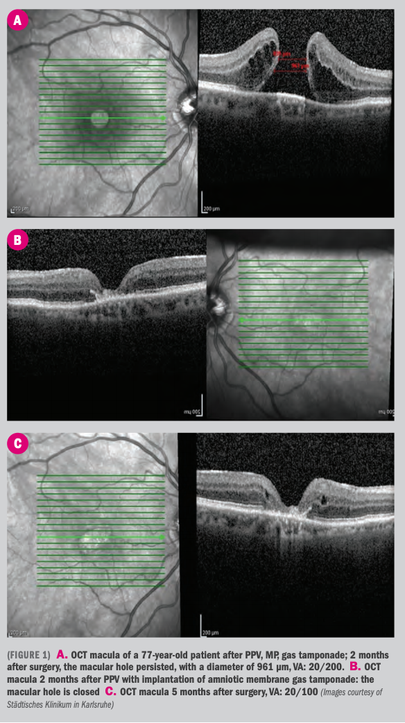
A novel, less invasive alternative to autologous tissue transplantation in the surgical treatment of large and recurrent MHs is the use of human amniotic membrane (hAM) transplantation (or implantation). With this method, the lens capsule and the peripheral retina of the affected eye are left intact. A plug of lyophilised or cryopreserved hAM is used instead of the patients’ tissues to repair the MH.
Use of hAM in medicine is not new. Applications of hAM in medicine in general, and in surgical ophthalmology in particular, have been reviewed.13 Until 2018, the use of hAM in clinical ophthalmology had been confined to the ocular surface.
In one study published in 2018, hAM patches were placed via an intraocular approach to repair large and recurrent MHs.14 Interestingly, from a historical point of view, repair of MHs with hAM had already been attempted in the mid 20th century, with a surgically complicated retro-bulbar approach in 1957 and in 1964.15,16
Animal and in vitro experiments preceded present day clinical use of hAM. Experiments carried out in pigs’ eyes showed the effect of transplanted amniotic membrane on subretinal wound healing.
Amniotic membrane modified choroidal neovascularisation after mechanical damage to Bruch’s membrane and seemed to behave as a basement membrane substitute for the proliferation of retinal pigment epithelium (RPE).17 In vitro, it was demonstrated that RPE cells cultured on hAM had an epithelial phenotype and secreted growth factors essential for retinal homeostasis.18
A 2018, prospective, consecutive case-series described positive results when hAM was implanted in eight patients who had large recurrent MHs. All patients had already undergone pars plana vitrectomy with ILM peeling and gas tamponade. The hAM was delivered cryopreserved from a human tissue bank and was defrosted intraoperatively before insertion.
In all patients, OCT at 1 week postoperatively showed MH closure with neurosensory retina overfilling the hAM. Best corrected visual acuity improved from 1.48±0.49 logarithm of the minimal angle of resolution (logMAR), (20/800) preoperatively to 0.71±0.37 logMAR (20/100) 3 months postoperatively, and to 0.48±0.14 logMAR (20/50) 6 months after the procedure. No ophthalmological adverse events were seen during follow-up.14
In a further study, hAM was used in ten patients with high myopia and MHs associated with retinal detachment who had undergone at least one pars plana vitrectomy. Half of the patients received silicon oil and half 10% octafluoropropane as tamponade at the end of surgery.
Silicon oil was removed 2 months after surgery. Results were very satisfying since retinal re-attachment
was achieved in all patients and visual acuity improved from 1.73 logMAR to 0.94 logMAR after 6 months.19
It appears that hAM is well tolerated. The possibility of hAM rejection has also been considered. In 1999, subretinal implantation of hAM in a rabbit model caused no evidence of inflammation or rejection.20
In the wake of these results, we started implanting amniotic membranes in patients who had large or recurrent MHs. However, a few of the steps in our surgical approach differs from that described by the aforementioned study: like them, we use a 23-gauge access but unlike their approach, we do not use a chandelier, so the method is not bimanual. The amniotic membrane is managed with a crocodile forceps.
A partial fluid–air exchange takes place, leaving a minimal amount of fluid at the foveal level in order to facilitate the manoeuvre of insertion of the amniotic plug. Implantation of the membrane may be facilitated by the use of a Tano scraper. No perfluorocarbon (PFCL) is used. Surgery is completed with fluid–air exchange and, finally, washout with perfluoropropane takes place.
The best possible outcome, in terms of anatomical results as well as visual acuity, is achieved when patients are able to remain in a facedown position for several days. In those who have physical disabilities or high comorbidities and who are, therefore, unable to hold the facedown position for a long time, silicon oil is used to keep the amniotic membrane in place after insertion.
Conclusions
Clinical studies are currently underway to confirm the promising results obtained so far with subretinal implantation of hAM plugs in the treatment of large and recurrent MHs. It seems that, in addition to anatomical success, the amniotic membrane stimulates retinal ingrowth and leads to improvements in visual acuity.
For this reason, we think attempts should be made to further the understanding of this novel technique in order to analyse its capability to restore visual function. We would suggest an accurate comparison of preoperative and postoperative measured parameters such as visual field assessment and electroretinogram exams, which can validate this technique.
In conclusion, we would affirm that the implantation of amniotic membrane may offer new hope in restoring vision in patients with an otherwise grim outlook.
Disclosures:
Emiliano Di Carlo, MD
e: emi.dicarlo@hotmail.it
Dr Di Carlo is a vitreoretinal consultant at the ophthalmology department of the Städtisches Klinikum Karlsruhe, Germany. His main interests are micropulse laser and new frontiers of vitreoretinal surgery. He has no financial interests in the subject matter.
Camilla Simini, MD
e: camilla.9128@gmail.com
Dr Simini is an ophthalmology resident at Städtisches Klinikum Karlsruhe, Germany, which is directed by Prof. A.J. Augustin. Her main interests are: medical and surgical retina. She has no financial disclosures.
References:
1. Knapp, H. Ãber isolierte Zerreissungen der Aderhaut infolge von Traumen auf dem Augenapfel. Arch Augenheilkunde. 1869;1:6-29.
2. Aaberg, TM. The macular holes. A Review. Surv Ophthalmol. 1970;15:139-162.
3. Stalmans P, Benz M, Gandorfer A et al. Enzymatic vitreolysis with ocriplasmin for vitreo-macular traction and macular holes. New Engl J Med. 2012;367:606-615.
4. Kelly NE, Wendel RT. Vitreous surgery for idiopathic macular holes. Results of a pilot study. Arch Ophthalmol. 1991;109:654-659.
5. Tadayoni R, et al. Relationship between macular hole size and the potential benefit of internal limiting membrane peeling. Br J Ophthalmol. 2006;90:1239-1241.
6. Ullrich,et al. Macular hole size as a prognostic factor in macular hole surgery. Br J Ophthalmol. 2002;86:390-393.
7. Michalewska, et al. Inverted internal limiting membrane flap technique for large macular holes. Ophthalmology. 2010;117:2018-2025.
8. Michalewska, et al. Temporal inverted internal limiting membrane flap technique versus classic inverted internal limiting membrane flap technique: a comparative study. Retina. 2015;35:1844-1850.
9. Velez-Montoya R, et al. Inverted ILM flap, free ILM flap and conventional ILM peeling for large macular holes. Int J Retina Vitreous. 2018;19:8.
10. Purtskhvanidze K, et al. Persistent full-thickness idiopathic macular hole: anatomical and functional outcome of revitrectomy with autologous platelet concentrate or autologous whole blood. Ophthalmologica. 2017;239;1:19-26
11. Chen SN, et al. Lens capsular flap transplantation in the management of refractory macular hole from multiple etiologies. Retina. 2016;36:163-170.
12. Grewal, et al. Autologous neurosensory retinal free flap for closure of refractory myopic macular holes. JAMA Ophthalmol. 2016;134:229-230.
13. Murube J. Early clinical use of amniotic membrane in medicine and ophthalmology. The Ocular Surface. 2006;4:114-119.
14. Rizzo, et al. A human amniotic membrane plug to promote retinal breaks repair and recurrent macular hole closure. Retina. 2018. doi.org/10.1097/
IAE.0000000000002320.
15. Csapody I. Amnion Implantation gegen Maculaloch (Amniotic membrane implantation against macular holes). Ophthalmologica, 1957;134:273-275.
16. Bechrakis E, Soellner F. Zur Amnion Implantation bei Makulaforamen [On amnion implantation in macular foramina.] Ber Zusammenkunft Dtsch Ophthalmol Ges. 1964;65:81-83.
17. Kiilgaard JF, et al. Transplantation of amniotic membrane to the subretinal space in pigs. Stem Cells Int, 2012. Doi. org/10.1155/2012/716968.
18. Ohno-Matsui K, et al. The effects of amniotic membrane on retinal pigment epithelial cell differentiation. Mol Vis. 2005;6:1-10.
19. Caporossi T, et al. A human Amniotic Membrane plug to manage high myopic macular hole associated with retinal detachment. Acta Ophthalmol. 2019.doi. org/10.1111/aos.14174
20. Rosenfeld PJ, et al. Subretinal implantation of human amniotic membrane: a rabbit model for the replacement of Bruch’s membrane during submacular surgery. Invest Ophthalmol Vis Sci. 1999;40:206.
Newsletter
Keep your retina practice on the forefront—subscribe for expert analysis and emerging trends in retinal disease management.