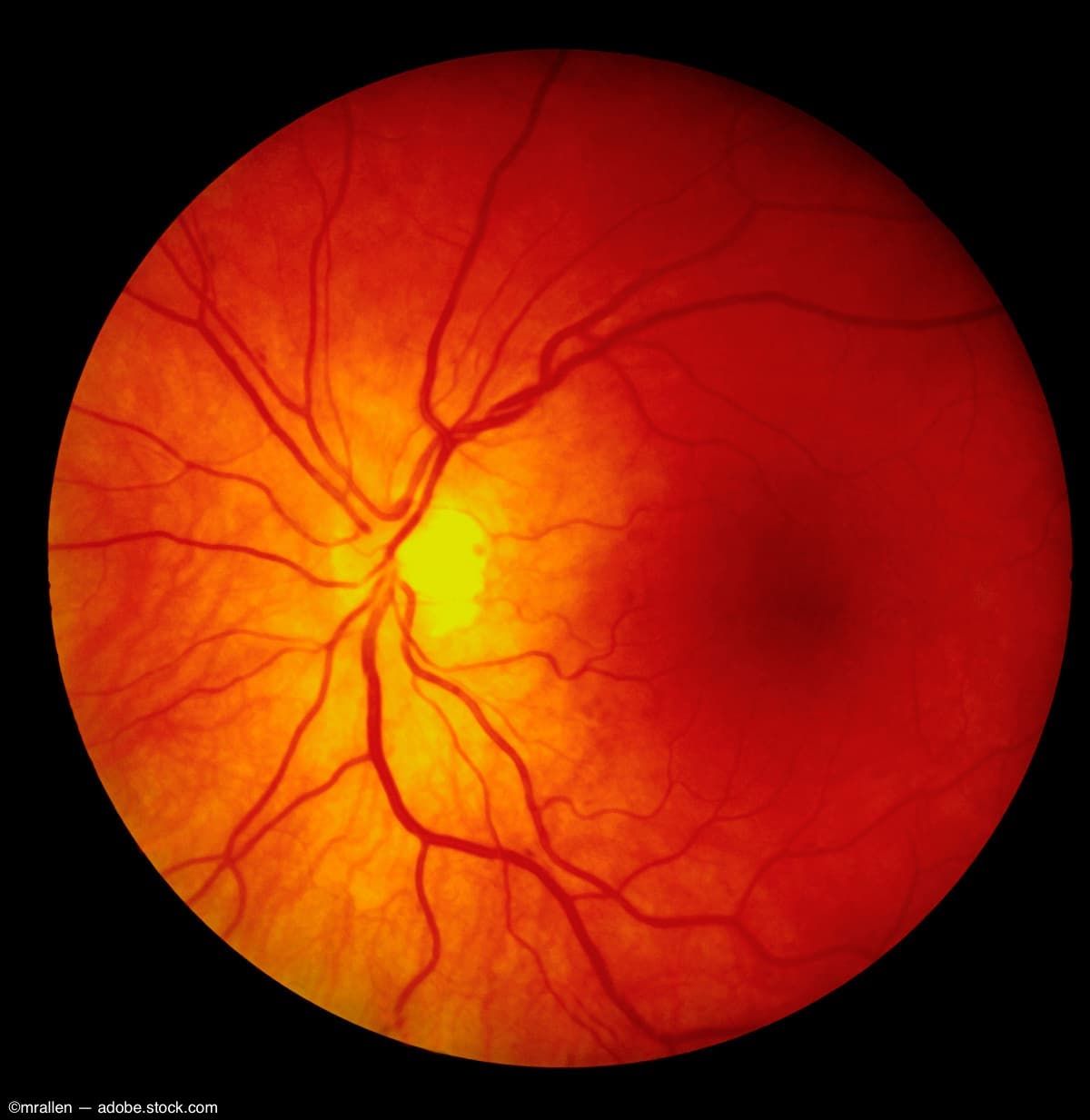
Structural and vascular changes detected in RVO affected eyes and unaffected fellow eyes
(Image credit: AdobeStock/mrallen)

Both eyes of patients with retinal vein occlusion (RVO), regardless of whether the eye is affected or is the unaffected fellow eye, have central and/or peripheral structural and vascular alterations in the retina and choroid,1 reported Xin-Yu Zhao, PhD, and colleagues.
Zhao is from the Department of Ophthalmology, Peking Union Medical College Hospital, Chinese Academy of Medical Sciences, and the Key Laboratory of Ocular Fundus Diseases, Chinese Academy of Medical Sciences & Peking Union Medical College, Beijing, China.
The investigators cited numerous gaps in knowledge that include poorly characterized RVO-associated choroidal changes, with conflicting findings regarding subfoveal choroidal thickness (SFCT) and flow density in the choriocapillaris.2,3; a lack of information about changes in the large and medium choroidal vessels; and Incompletely studied pathologic alterations in the fellow eyes of patients with RVO despite systemic vascular associations of RVO that likely impact the fellow eye, but the fellow eyes were never compared to healthy control eyes.4
In addition, the two-dimensional choroidal vascularity index (CVI), ie, the ratio of the choroidal luminal area to the total choroidal area in 1 optical coherence tomography (OCT) B-scan, that can quantitatively analyze the choroidal structure in various retinal diseases,5 was replaced by the three-dimensional CVI assessment using the entire OCT volume, but this latest iteration of the assessment has not been evaluated in RVO.
They also mentioned that ultra-widefield (UWF) swept-source OCT angiography (OCTA) (24 x 20 mm) used in this study provides a wider field of view compared with the limited 3 x 3 to 12 x 12 fields used, and facilitated evaluation of regions beyond the posterior pole.
Prospective OCTA study
The investigators conducted a prospective UWF OCTA study to evaluate 15 patients with central RVO (CRVO) and 15 patients with branch RVO (BRVO) and compared the findings to those of 15 healthy age-matched controls.
The retinal and choroidal parameters that were evaluated in the central and peripheral areas were the retinal vessel flow density (VFD) and vessel linear density (VLD), choroidal vascularity volume (CVV), CVI, and VFD in the large and medium choroidal vessels (LMCV-VFD).
The results indicated that eyes with ischemic CRVO and BRVO had an increased foveal avascular zone area, perimeter, and acircularity index compared to fellow and healthy control eyes, and the fellow eyes of those with RVO also had larger acircularity index values than controls (P < 0.05).
When eyes with ischemic CRVO and BRVO were compared with control eyes, the results showed that the VFD, VLD, CVV, CVI, and LMCV-VFD decreased, but the retinal thickness and volume in the superficial capillary plexus, deep capillary plexus, and whole retina increased (P < 0.05).
In addition, the fellow eyes of those with RVO also had significantly decreased retinal VFD, LMCV-VFD, and CVI and increased retinal thickness and volume compared with control eyes (P < 0.05). The alterations were inconsistent throughout the retina, and in some cases involved only the peripheral or central regions.
The authors concluded, “The affected and unaffected fellow eyes of RVO patients showed both central and/or peripheral structural and vascular changes affecting the retina and choroid. UWF-OCTA, which enables visualization of the more peripheral fundus regions, offers a promising approach to more fully characterize vascular alterations in RVO.”
References
1. Zhao X-y, Zhao Q, Wang C-t, et al. Central and peripheral changes in retinal vein occlusion and fellow eyes in ultra-widefield optical coherence tomography angiography. Invest Ophthalmol Vis Sci. 2024;65:6; doi:https://doi.org/10.1167/iovs.65.2.6
2. Khodabandeh A, Shahraki K, Roohipoor R, et al. Quantitative measurement of vascular density and flow using optical coherence tomography angiography (OCTA) in patients with central retinal vein occlusion: can OCTA help in distinguishing ischemic from non-ischemic type? Int J Retina Vitreous. 2018;4:47.
3. Rayess N, Rahimy E, Ying GS, et al. Baseline choroidal thickness as a predictor for treatment outcomes in central retinal vein occlusion. Am J Ophthalmol. 2016;171:47–52.
4. Shin YI, Nam KY, Lee SE, et al. Changes in peripapillary microvasculature and retinal thickness in the fellow eyes of patients with unilateral retinal vein occlusion: an OCTA study. Invest Ophthalmol Vis Sci. 2019; 60: 823–829.
5. Hwang BE, Kim M, Park YH. Role of the choroidal vascularity index in branch retinal vein occlusion (BRVO) with macular edema. PLoS One. 2021;16:e0258728.
Newsletter
Keep your retina practice on the forefront—subscribe for expert analysis and emerging trends in retinal disease management.