Artificial retinal device increases edge of contrast, mimics human optical illusions
Investigators at the International Center for Materials Nanoarchitectonics have developed the first-ever artificial retinal device that increases the edge contrast between lighter and darker areas of an image, using ionic migration and interaction within solid.

A team at the International Center for Materials Nanoarchitectonics (WPI-MANA) has developed the first-ever artificial retinal device that increases the edge contrast between lighter and darker areas of an image, using ionic migration and interaction within solid.
According to a WPI-MANA news release, the device has the potential for use in developing compact, energy-efficient visual sensing and image-processing hardware systems capable of processing analog signals.
Recently, artificial intelligence (AI) system developers have shown much interest in research on various sensors and analog information-processing systems inspired by the human senses. Most AI systems require sophisticated software and complex circuit configurations, including custom-designed processing modules. The problem with these systems is that they are large and consume much power.
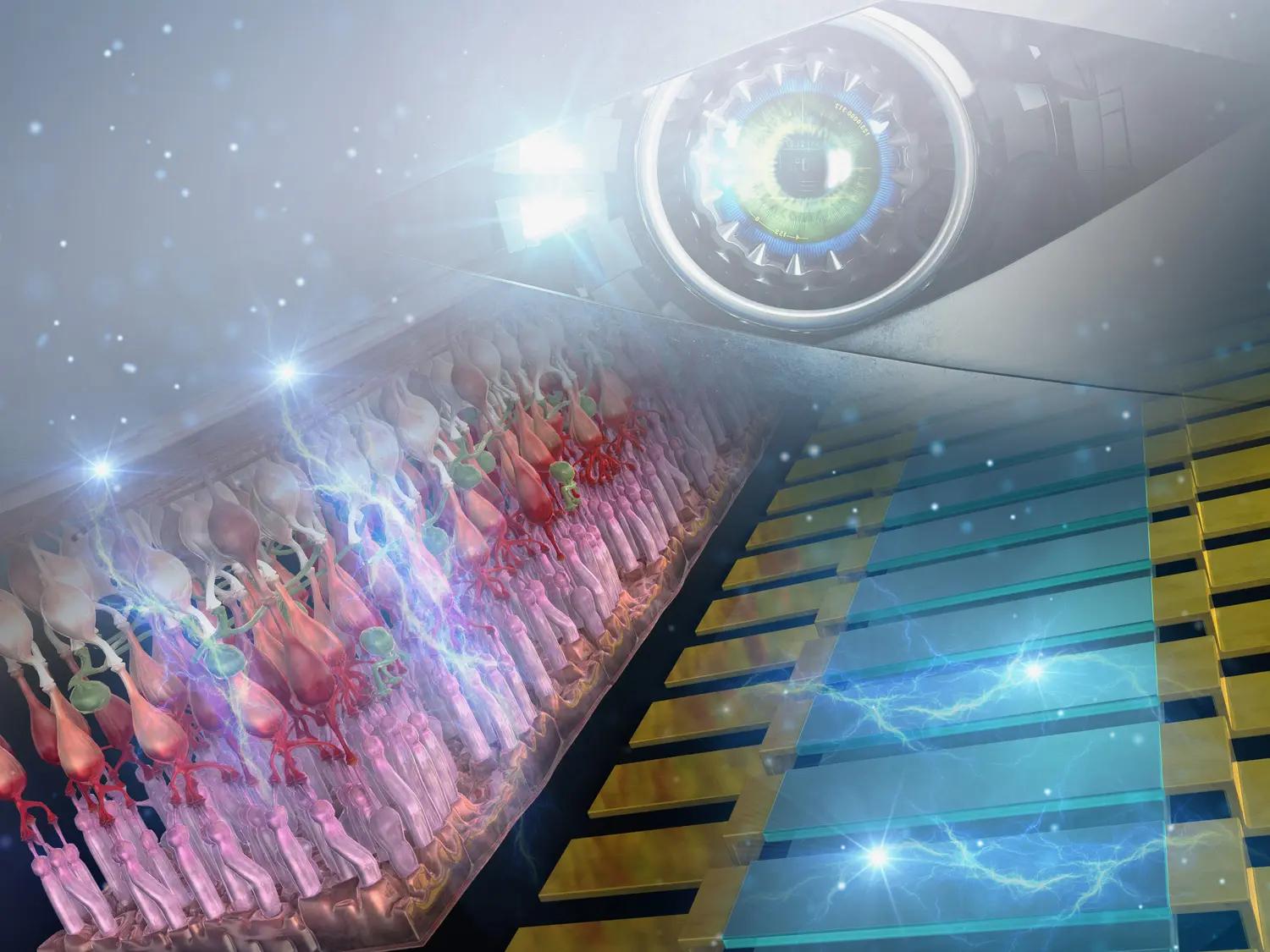
Artificial intelligence (AI) system developers have shown much interest in research on various sensors and analog information-processing systems inspired by the human senses. (Image courtesy of the International Center for Materials Nanoarchitectonics)

According to the center, the team built a multiple ionic device system, each of which had a lithium cobalt oxide channel arranged on a common lithium phosphorus oxynitride electrolyte. Because of the migration of Li-ions between the channels through the electrolyte, the devices were highly interactive, similar to human retinal neurons such as photoreceptors, and horizontal and bipolar cells. Input voltage pulses caused ions within the electrolyte to migrate across the channels, which changed the output channel current.
The center, which is located in Ibaraki, Japan, noted that the device was able to process input image signals and produce an image with increased edge contrast between darker and lighter areas. The center also noted that this is similar to the human visual system's ability to increase edge contrast between brightness differences by means of visual lateral inhibition.
According to investigators at WPI-MANA, the human eye produces various optical illusions associated with tilt angle, size, color and movement, in addition to darkness/lightness, and this process is believed to play a crucial role in the visual identification of different objects.
The center pointed out that the artificial retinal device the team created could be used to reproduce these types of optical illusions. They hope to develop visual sensing systems capable of performing human retinal functions by integrating their device with other components, including photoreceptor circuits.
This research was conducted by Tohru Tsuruoka, a chief researcher at Nanoionic Devices Group, WPI-MANA, NIMS; Kazuya Terabe, MANA principal investigator and group leader, Nanoionic Devices Group, WPI-MANA, NIMSand their collaborator.
Reference
Xiang Wan, Tohru Tsuruoka, Kazuya Terabe; Neuromorphic System for Edge Information Encoding: Emulating Retinal Center-Surround Antagonism by Li-Ion-Mediated Highly Interactive Devices. Nano Letters 2021, 21, 19, 7938-7945; Published Sept. 13, 2021; DOI: 10.1021/acs.nanolett.1c01990
Newsletter
Keep your retina practice on the forefront—subscribe for expert analysis and emerging trends in retinal disease management.