- Modern Retina Fall 2024
- Volume 4
- Issue 3
Spotlight on GA: Promoting early detection and treatment
Murtaza K. Adam, MD, adjunct clinical associate professor at Rocky Vista University in Parker, Colorado, and chair of clinical research at Colorado Retina in Denver, Colorado, described his approach to treating patients with geographic atrophy (GA), current therapies, and safety concerns during a Modern Retina
Managing GA
Management of this population has changed. Previous examinations were characterized by sympathy and advising patients to continue AREDS vitamins (Bausch + Lomb) and a Mediterranean diet, as well as active treatment with the introduction of 2 complement inhibitors, avacincaptad pegol intravitreal solution (Izervay; Astellas) and pegcetacoplan injection (Syfovre; Apellis Pharmaceuticals), that can slow GA progression and delay functional vision loss.
Adam initially focused treatment with these drugs on patients with vision loss in 1 eye and relatively good visual acuity (VA) in the other. “They were highly motivated to begin treating GA to slow its progression,” Adam said.
It is important to be mindful of the adverse effects associated with complement inhibitors. There is a slightly higher risk of neovascular age-related macular degeneration (AMD) development compared with patients not treated with complement inhibitors. Occlusive retinal vasculitis is another risk, although low, that can cause extremely debilitating and irreversible vision loss. Others include endophthalmitis, retinal detachment, and elevated IOP, he explained.
Adam’s approach is to begin a trial of complement inhibitors in the worse-seeing eye to monitor for adverse effects to evaluate the patient’s risk profile; he never uses complement inhibitors in patients with a history of uveitis or an inflammatory response to another drug. He also said that he does not begin treatment with complement inhibition therapy on the first visit because of the importance of monitoring for GA progression over time, the risk of vision loss in some instances, and ensuring that the patient is well educated on the risks and benefits of treatment.
Patients with GA are heterogeneous (ie, some have non–center-involving or center-involving GA and concurrent neovascular AMD). Those with non–center-involving GA might be observed for 6 to 8 monitoring sessions to determine the rate of change over time.
Case 1
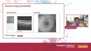
Fundus imaging of the left eye showed a similar scenario with a great deal of calcific drusen in the macula and posterior pole. Multimodal autofluorescence imaging more readily detected GA, highlighting the high vision loss risk. A nasal, circumferential band of GA, multifocal in nature, was seen; at the GA edge, the hyperautofluorescence was concerning for a high risk of progression. OCT showed a loss of the RPE layer and the intersegment outer segment junction nasal to the foveal center. Education about the risks and benefits of treatment was important for this patient, who was instructed about the slightly higher risk of neovascular AMD associated with complement inhibitors and retinal vasculitis.
The patient was treated with pegcetacoplan every 6 to 8 weeks. The FDA label for pegcetacoplan permits monthly or every-other-month treatment. “In most cases, I recommend treatment every 6 to 8 weeks because that timing rides the line between treatment efficacy and the risks associated with frequent complement inhibitor therapy,” he explained. The patient had been treated for about 1 year with no treatment-related adverse events.
Case 2
Examination of the left eye showed calcific drusen throughout the macula and multifocal non–center-involving GA threatening the fovea. There was a hyperfluorescent pattern at the edges of some of the GA.
Because this patient was legally blind in her right eye, it was essential to protect her left eye. In this situation, atrial injection of complement treatment was tested in the right eye before starting treatment in the other eye, and she was evaluated 4 to 6 weeks later. The symptom onset, according to Adam, begins 10 to 14 days after injection, but that did not occur in this patient in the left eye.
In this case, treatment in the left eye was scheduled for 4- to 6-week intervals because of concern about the timeline for vision loss in that eye. Choroidal neovascularization (CNV) developed about 4 months later in that eye. Complement therapy was stopped, and anti-VEGF therapy was started with aflibercept (Eylea; Regeneron) on a treat-and-extend regimen of about 14 months. The left eye vision remained stable at 20/30. The right eye’s vision improved from 20/400 to 20/100 following cataract surgery despite a large area of center-involving GA.
Case 3
The first examination showed that the right eye had intermediate AMD; the left had more advanced AMD with a non–center-involving area of GA temporal and superior to the fovea. The left eye was observed initially because no complement inhibition therapies were available. At 8 months, she reported decreased contrast sensitivity and night vision despite the stable VA. Infrared imaging showed significantly more GA, and at 16 months, this process continued.
She returned to the clinic at 16 months, following extensive European travel where complement inhibitors were unavailable. The left eye vision declined from 20/50 to count fingers, with a great deal of GA around the fovea. OCT showed an area of subretinal fibrosis, indicating a possible subretinal hemorrhage due to CNV, which resulted in central loss of photoreceptors and RPE. The GA developed slowly in her right eye compared with the left eye. A non–foveal-sparing area of GA superotemporal to the fovea and another small area inferiorly were seen. Compared with baseline, this patient’s risk for central visual loss increased over time.
Adam considered complement inhibition therapy for this patient. Following a discussion with the patient, a trial injection of avacincaptad pegol was administered to the left eye to rule out adverse effects. A dilated fundus examination 6 to 8 weeks later ruled out intraocular inflammation treatment in her right eye, and the treatment continued but with numerous travel interruptions.
The obvious concern was that a lack of consistency with this therapy would lead to more rapid vision loss. Adam concluded, “Because of the numerous factors that can interfere with timely treatment, my hope is that we’re still chipping away at the progression of GA with the treatments that we are able to provide.” •
Reference
Adam M. Spotlight on Geographic Atrophy: Promoting Early Detection and Treatment. Modern Retina. July 18, 2024. Accessed August 14, 2024. https://www.modernretina.com/case-based-roundtable-series/spotlight-on-geographic-atrophy-promoting-early-detection-and-treatment
Articles in this issue
about 1 year ago
A great leap forward in ophthalmic drug delivery?about 1 year ago
The sights of Stockholmabout 1 year ago
Higher molar dose in the real world and clinical trialsover 1 year ago
The unique role of the retina optometristover 1 year ago
The future of retina careNewsletter
Keep your retina practice on the forefront—subscribe for expert analysis and emerging trends in retinal disease management.


