Anti-Vascular Endothelial Growth Factor (Anti-VEGF) Therapies in Neovascular AMD and DME
Adrienne Scott, MD; Carl Regillo, MD, FACS, FASRS; and Prethy Rao, MD, MPH, provide an overview of the effect of anti-VEGF treatment in neovascular age-related macular degeneration and diabetic macular edema.
Episodes in this series

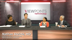
Nathan Steinle, MD: Hello, and welcome to this Modern RetinaTM Viewpoints titled, “Enhancing Vision Outcomes with Innovative Therapies in Neovascular AMD and DME.” I am Dr Nathan Steinle, a vitreoretinal specialist at California Retina Consultants in Santa Barbara, California. Joining me today in this discussion are Dr Adrienne Scott, a vitreoretinal surgeon at Wilmer Eye Institute, Johns Hopkins University School of Medicine in Baltimore, Maryland; Dr Carl Regillo, chief of retina services at Wills Eye Hospital in Philadelphia, Pennsylvania; and Dr Prethy Rao, a retina specialist, Retina and Vitreous Associates of Texas in Houston, Texas.
Our discussion today will focus on the latest therapies for neovascular AMD [age-related macular degeneration] and DME [diabetic macular edema] including their efficacy, safety, and durability. Additionally, we will share evidence-based strategies and practical tools for improving outcomes in patients with these retina diseases. Welcome and let’s get started.
Let me throw out first to you Dr Scott, that these anti-VEGF agents have been around for almost 20 years. Talk to me a little bit about the history of the anti-VEGF area and how it revolutionized both AMD and DME.
Adrienne Scott, MD: Thank you Dr Steinle. I’m fortunate to have spent most of my career with these available to me and the anti-VEGF agents have definitely changed outcomes and expectations for our patients. They’re living longer and being able to enjoy more quality of life-related to their vision. Corneal neovascularization used to be a sentence for legal blindness for patients often and even in diabetic macular edema and diabetic eye disease, the number of treatments we have increased so dramatically. We used to just have a laser to modify the disease process and maybe stabilize vision, prevent severe vision loss, but now the expectations for both the doctor and patient are not only vision stabilization but improves vision so it’s a great time to be a retina specialist.
Nathan Steinle, MD: Absolutely. So, Dr Rao, I’m going to throw it to you next. We know that these injections work well but it’s also a significant burden for the patient and their families. Talk to me a little bit about how you are trying to reduce burdens in your practice and how you perhaps do treat and extend.
Prethy Rao, MD, MPH: Great question, Nathan. The first thing I do is have a discussion with the patient and family about what their needs are. I discuss how far they live from the clinic in terms of transportation because that really affects if they’re able to make their visit. I do a needs assessment in terms of what their resources are.
The second thing I do in terms of how I treat patients is I’m a quick extender. I usually start with 3 injections in a row monthly for 3 months, but if after the second injection they’re doing very well and I don’t see any active fluid and their vision plateaus then I do extend them out pretty quickly, 1- to 2-week regimens. That’s what I start out with.
Nathan Steinle, MD: Fantastic. Dr Regillo, so I’m lucky that I remember my old fluorescein days. We used to have fluorescein conferences. I’d get grilled and now this beautiful tool of OCT [optical coherence tomography] is out. Talk to me about what you use in your clinics. You guys are at the pinnacle of eye care there at Wills Eye Hospital. What do you do in your typical patient visits for imaging?
Carl Regillo, MD, FACS, FASRS: Well, it’s very true that at one point in the very distant past, fluorescein angiogram was the gold standard to diagnose neovascular AMD and, in some respects, as you mentioned, guiding treatment. That changed in the other revolution that took place. In fact, right around the time, anti-VEGFs hit the scene, OCT was becoming a very common and now an essential tool and the imaging primary mode to diagnose and guide treatment with anti-VEGF therapy. No doubt about it. We really couldn’t live without it and if we did, we’d have to put everyone on a monthly injection regimen or every other month. With it, we’re able to individualize treatment for both neovascular AMD and our other retinal conditions.
Nathan Steinle, MD: Do you image a person at the OCT every single visit?
Carl Regillo, MD, FACS, FASRS: Absolutely, and not only the eye of interest, the eye we’re treating, but the fellow eye. This is a bilateral disease and our patients with wet AMD in 1 eye are at very high risk for developing wet AMD, going from dry to wet in the fellow eye. Within a 5-year time frame, that could be 50% or greater so we get our best results with these drugs when this disease is caught early. Imaging the fellow eye at every visit in addition to the eye we’re treating, of course, allows us to pick up on that wet AMD transformation at the very earliest stages.
Transcript edited for clarity
Newsletter
Keep your retina practice on the forefront—subscribe for expert analysis and emerging trends in retinal disease management.

