Biomarkers of Disease at Baseline in Neovascular AMD
Retina experts discuss biomarkers to track disease progression in neovascular AMD, as well as their thoughts on the utility of optical coherence tomography angiography (OCT-A).
Episodes in this series

Nathan Steinle, MD: I’ll go back to 1 more question on neovascular AMD [age-related macular degeneration], and it’s 1 I struggle with. Patients ask me, “How many treatments am I going to get? What’s my burden going to look like?” I’m a poor predictor. I look at the OCT [optical coherence tomography]. I look at the FA [fluorescein angiography]. What biomarkers do you look at, at baseline, that give this patient’s information and what their disease course is going to look like? Dr Scott?
Adrienne Scott, MD: It’s so variable among patients. Even patients with choroidal neovascularization and wet AMD can be quite heterogeneous. If somebody has subretinal hemorrhage present, that’s a patient I’d like to be very aggressive with. Those are the patients I want to see resolution of the subretinal hemorrhage before I start extending. That’s 1 of the biggest predictors of active disease that we can see and monitor. Those patients have worse prognoses, so I want to be aggressive with their disease. For patients who seem to get dry very quickly after 1 or 2 injections, I feel a bit more hopeful. I try to extend them. I might extend them a little more rapidly.
Nathan Steinle, MD: How about this side of the table? What biomarkers do you look at, at baseline?
Prethy Rao, MD, MPH: I agree 100%. I’d say subretinal hemorrhage. The presence of hemorrhage often predicts it. I usually hesitate to give them a number of injections. I say, “The clinical trials and real-world studies say 6 to 8 in the first year, but that might change.” I usually say, “Let’s see how things go after 1 or 2, and then I’ll be able to better predict how you might end up needing the number of injections.”
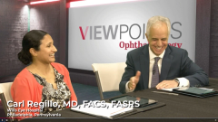
Carl Regillo, MD, FACS, FASRS: I have a similar dialogue with the patient. This is the nuance for a patient with wet AMD. At baseline, you can’t predict the need or the frequency of injections with any of these drugs. You can, however, see baseline features that predict vision outcomes. If you caught the disease early, your vision is good, and your lesion is small, that has a better prognosis for a better vision outcome. That’s what you can say. You may not want to provide that perspective because it’s such a variable response. I tell the patient we’re going to start treating monthly until everything is as good as possible.
Then we’ll start to extend the treatments slowly if we can. Everyone is different, even though eventually that patient often falls into a certain fixed pattern. They might ultimately spend 8 weeks on drug X, Z, or whatever, or 10 weeks. They’re pretty consistent in that way, but even that can change over time.
Adrienne Scott, MD: Here’s where I like to employ OCT as a teaching tool in the office with the family, so they understand what I’m looking for and why I’m recommending a certain follow-up interval. We all look at the skin, and I say, “It’s a collaborative approach. We’re going to talk it out and look at it.” I show them what fluid is. They know what fluid looks like. They know and understand this is more effective vs not as effective based on the presence or absence of that fluid. It’s a nice way to collaborate with the patients, and they understand why you’re recommending that follow-up interval. Also, I’m clear with them that it’s hard to predict what will happen. This is going to be a lifelong journey. You might get to the point where you don’t need as many injections, but we’ll determine that down the line.
Nathan Steinle, MD: Great points. I’m going to dovetail off that to OCT angiography [OCT-A]. We have OCT in all our clinics. I’m not utilizing it as much as I thought I would when the technology came out 5 years ago. Do you use OCT every day? If so, teach me how you do it.
Adrienne Scott, MD: I’m not as fast with the OCT angiography technology and these quick decision-making scenarios about what you’re deciding to treat. It’s great if you’re not sure if there’s an agitative CNV [choroidal neovascularization] going on. Sometimes, patients will have these subfoveal or vitelliform lesions, and you’re not sure if there’s a choroidal neovascular lesion there. You also have to think about the reimbursement aspect of it because you can’t get reimbursed for an OCT and an OCT angiography study on the same day. I use it, but it’s more of an academic thing when I look and I want to see if there’s a CNV active lesion, but I’m not using it every day in decision-making—only in select cases.
Nathan Steinle, MD: How about this side? OCT-A, yes or no?
Carl Regillo, MD, FACS, FASRS: It’s not part of routine retina care. It’s a very intriguing, interesting technology. It has a role in clinical practice, in differentiating neovascular AMD from disease that looks like it. Up-front diagnosis can help you. Is this central serous or neovascular AMD? Once you know what you’re dealing with, once it’s neovascular AMD, it adds little if anything to the management going forward. You can treat and manage neovascular AMD to the highest standard without OCT-A.
There’s no study that says OCT-A affects the outcomes in any way. It’s cumbersome and not easy to use, and the images often make you scratch your head as you try to figure out what’s going on here. It’s not a perfect technology, but it’s very promising. Even now, there’s some use in the clinical setting.
Prethy Rao, MD, MPH: I agree 100%. For the burden and the time it takes to give OCT-A, for some of our older patients who aren’t able to sit in place for a while, it becomes challenging. It would be interesting to see how OCT-A plays a role in a lot of these extended patients—3, 4, or 5 months. Do they end up getting to that period but still have an active CNV lesion? For some of these patients that we get to the longer intervals when they recur, we use OCT-A to see if their CNV is still active to modify or personalize their care. But for day-to-day practice, I agree with Drs Regillo and Scott that it’s hard to do.
Carl Regillo, MD, FACS, FASRS: You brought up a good point earlier. For neovascular AMD, we’re not treating just the wetness, the exudative manifestations. We’re also stopping the growth of neovascularization, even from the early studies in which fluorescein angiography was used at baseline and the final follow-up. On average, CNV lesions did not grow. CNV can grow with relative undertreatment, even with few or no signs of exudation. That can be detrimental and could account for some of the decreased vision over time. Growing lesions can be invasive. You don’t want to see CNV grow large, for the most part. There could be exceptions, depending on the level of the CNV, where it’s located, and how destructive it is. But I’ve seen people lose vision based on CNV growth with little to no exudation, not just with neovascular AMD but also myopic CNV.
Nathan Steinle, MD: I’m curious if our use of OCT-A will pick up as we treat more patients with dry AMD. That conversion from dry to wet is interesting.
Transcript edited for clarity
Newsletter
Keep your retina practice on the forefront—subscribe for expert analysis and emerging trends in retinal disease management.


