- Modern Retina Summer 2021
- Volume 1
- Issue 1
Laser imaging system may offer early detection, treatment for eye diseases that cause blindness
According to investigators, the new technology is designed to detect telltale signs of major blinding diseases in retinal blood and tissue that typically go unseen until it is too late.
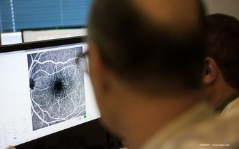
Investigators believe a non-contact laser imaging system could help doctors diagnose and treat eye diseases that cause blindness much earlier than is now possible.
The new technology,1 developed by engineering investigators at the University of Waterloo in Waterloo, Ontario, Canada, is designed to detect telltale signs of major blinding diseases in retinal blood and tissue that typically go unseen until it is too late.
With current testing methods, diseases such as age-related macular degeneration, diabetic retinopathy and glaucoma, which have no symptoms in their early stages, are usually diagnosed only after vision is irreversibly affected.
“We are optimistic that our technology, by providing functional details of the eye such as oxygen saturation and oxygen metabolism, may be able to play a critical role in early diagnosis and management of these blinding diseases,” said Parsin Haji Reza, director of the PhotoMedicine Labs at Waterloo University, in a statement.
According to investigators, patented technology at the core of the new system is known as photoacoustic remote sensing (PARS). It uses multicolored lasers to almost instantly image human tissue without touching it.
When used for eyes, the non-invasive, non-contact approach improves both patient comfort and the accuracy of test results, according to the university.
The technology is also being applied by Haji Reza and investigators in his lab to provide microscopic analyses of breast, gastroenterological, skin and other cancerous tissues, and to enable real-time imaging to guide surgeons during the removal of brain tumors.
According to Richard Weinstein, MD, an ophthalmologist and co-founder of the Ocular Health Centre, PARS may move ophthalmology beyond the current gold standard in ophthalmological imaging.
“For the first time, not just in ophthalmology but in the entire medical field, diagnosis and treatment of disease could be made prior to structural change and functional loss,” he said in a statement.
Haji Reza, a professor of systems design engineering and co-founder of startup company illumiSonics, said researchers are working with several ophthalmologists and hope to start clinical trials within two years.
Reference
Z. Hosseinaee, N. Pellegrino, et. al; Functional and structural ophthalmic imaging using noncontact multimodal photoacoustic remote sensing microscopy and optical coherence tomography; Scientific Reports; May 2021; Accessed June 16, 2021.
Articles in this issue
over 4 years ago
Pediatric ophthalmologists target inherited retinal diseasesover 4 years ago
Gaining a greater understanding of AMDNewsletter
Keep your retina practice on the forefront—subscribe for expert analysis and emerging trends in retinal disease management.


