- Modern Retina Summer 2024
- Volume 4
- Issue 2
True-to-life retinal imaging with the new ultrawidefield color RGB modality
The introduction of red/green/blue (RGB) color is a welcome addition to ultrawidefield imaging that will aid in the screening and detection of retinal diseases.
Ultrawidefield (UWF) imaging is defined as retinal imaging beyond the vortex vein ampullae in all quadrants with a single capture, equating to a 110° to 220° view of the retina.1 Compared to the limited 30° to 50° view of traditional fundus photography, UWF imaging provides unparalleled access to the peripheral retina, enabling the visualization of previously undetected pathologies and deepening our understanding of retinal disorders.2
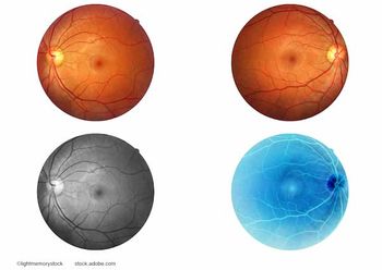
For example, the Optos UWF imaging device (Optos plc) Dunfermline, UK, is capable of capturing 200°, or 82% of the retina in a single image, which it achieves in less than 0.5 seconds without the need for mydriasis.3,4 Until recently, Optos UWF color imaging has relied on confocal scanning laser ophthalmoscopy with red (633 nm) and green (532 nm) lasers2; monochromatic red and green laser images are obtained, which are merged to form a pseudocolor retinal image.3 The addition of a blue laser (488 nm) to the Optos California device, one can obtain single-capture 200° red/green/blue (RGB) composite color retinal images that can mimic the natural color of the retina as seen on fundus examination.5,6
This new modality is a welcome feature, since it combines the extensive field of view of UWF with a natural retinal color representation. Color RGB images can be acquired simultaneously with color RG images, providing 2 UWF color images in 1 capture. The new modality also allows for blue laser autofluorescence in addition to green laser autofluorescence. Thus, more retinal detail can be obtained without increasing the test duration.
Promising avenues
Color RGB technology uses simultaneous red, green, and blue laser scans to acquire natural color images of the retina.5 Each of the laser sources in the RGB imaging system can also be applied individually to image different layers of the retina.4,7,8 While the green laser best visualizes the sensory retina and retinal pigment epithelium (RPE) and the red laser can image the choroid,8 the short-wavelength blue laser best captures the superficial retinal layers.7 Thus, in addition to color fundus photography, RGB imaging has potential application in the detection of retinal nerve fiber layer (RNFL) defects, which is a marker of early glaucomatous damage.9
Indeed, confocal scanning laser ophthalmoscopy with blue laser has been found useful in detecting RNFL defects in cases of suspected glaucoma.10
UWF RGB is also likely to be a useful diagnostic tool in other conditions where clinical fundus examination is precluded due to poor ocular fixation. UWF RG imaging has been able to successfully capture panoramic retinal images in patients with nystagmus,11,12 but RGB imaging can further expand the information we obtain from the acquired images.
An initial study investigating the clinical benefits of RGB color imaging reported clear visualization of vitreoretinal and superficial retinal structures, such as vitreous opacities and optic nerve fibers, due to the inclusion of blue laser.5 Pathologies such as epiretinal membrane and proliferative vitreoretinopathy were clearly visualized on RGB imaging. The high contrast of RGB images enhanced the visibility of peripheral retinal holes, tears, schitic changes, and lattice degenerations. The natural color demonstrated various retinal lesions, such as drusen, retinal exudates, superficial retinal hemorrhages, neovascularization, ghost vessels, and retinochoroidal atrophy. On the other hand, deep retinal or choroidal structures were better imaged with the RG modality, which improved visualization of subretinal fibrosis, retinal and choroidal vasculature, and deep retinal hemorrhages. RG images characterized RPE changes and enhanced the contrast between areas of geographic atrophy and normal retina in age-related macular degeneration (AMD). Thus, the findings of this study suggest that UWF RG and RGB imaging are complementary techniques that provide valuable information for more precise clinical decision-making.
Early clinical experience
At the Lariboisière Hospital, Paris, we were fortunate to get early access to the new UWF RGB imaging system.It was immediately evident that the
acquired images had enhanced clarity and more natural colors true to what is seen on clinical fundus examination, but our primary concern was to investigate whether these refinements improved our ability to diagnose and treat patients.
We found that minute retinal details were clearly visible, which is clinically significant in cases of diabetic retinopathy (DR) where undetected small hemorrhages and microaneurysms can lead to underestimation of true DR severity.3 We observed that color RGB replicated the actual appearance of the retina, making subtle hemorrhages and intraretinal microvascular abnormalities (IRMA) easy to spot (Figure 1). Crisp images were obtained even in cases with suboptimal viewing conditions due to media opacities. Thus, we could detect early lesions in addition to more peripheral lesions, enabling accurate staging and treatment of DR. In the initial results from our ongoing study comparing UWF RG and RGB imaging in DR, we were able to detect more hemorrhages and IRMA, as well as more cases of moderate and severe DR, with RGB images.
The high contrast and visibility of color RGB images were also expected to assist in the detection of peripheral retinal holes and tears. In the case shown in Figure 2a, a retinal detachment was noted on UWF RGB imaging with the eye in primary position. Although the UWF image showed the location of the detachment with a single, 200° retinal capture, it was not immediately apparent if there were any associated retinal tears. However, on changing the fixation point (gaze-steering), we were able to observe the far periphery, revealing tears in the superior and superonasal peripheral retina, which were clearly visible due to the natural color representation of RGB image (Figure 2b). A feature of the UWF software is its built-in auto-montage capability: The device automatically combines the gaze-steered images into a single image that captures 97% of the retina,⁴ showing the full extent of the detachment with all associated tears (Figure 3).
Figure 4 shows another display of the auto-montage feature in a patient post-retinal detachment surgery, where the panoramic color image makes it appear as if the entire eye has been laid open for the examiner.
In case of ocular tumors, an accurate representation of tumor appearance is essential for a precise diagnosis of the underlying pathology.13 With the RGB modality, we found that the color features of ocular tumors were faithfully imaged, enabling accurate evaluation of lesions. Color RGB was especially helpful in examining melanocytic lesions to distinguish choroidal nevi from choroidal melanomas and follow-up.
Other ophthalmologists have had similarly positive experiences with the new UWF RGB images, noting the great clarity and detail for a variety of retinal disorders.6
Conclusion
Early clinical experiences suggest that the addition of color RGB modality to UWF imaging is a new tool in our diagnostic toolbox that will be useful in accurately and efficiently diagnosing retinal pathologies, even in conditions unconducive to reliable retinography such as coexisting media opacities. This new modality is a significant addition in the field of UWF imaging that empowers physicians to provide more bespoke patient care. With its natural colors and expansive views of the retina, UWF RGB is likely to play an integral role in the diagnosis and management of retinopathy in routine ophthalmic practice.
Ali Erginay, MD
e: ali.erginay@aphp.fr
Erginay is senior consultant ophthalmologist at Lariboisière Hospital, Paris, France, and specializes in vitreoretinal disorders. The author sits on the Optos medical advisory board and has no financial interests to disclose.
References:
1. Choudhry N, Duker JS, Freund KB, et al. Classification and Guidelines for Widefield Imaging. Ophthalmol Retina. 2019;3(10):843-849. doi:10.1016/j.oret.2019.05.007
2. Kumar V, Surve A, Kumawat D, et al. Ultra-wide field retinal imaging: A wider clinical perspective. Indian J Ophthalmol. 2021;69(4):824. doi:10.4103/ijo.IJO_1403_20
3. Hirano T, Imai A, Kasamatsu H, Kakihara S, Toriyama Y, Murata T. Assessment of diabetic retinopathy using two ultra-wide-field fundus imaging systems, the Clarus® and OptosTM systems. BMC Ophthalmol. 2018;18(1):332. doi:10.1186/s12886-018-1011-z
4. Optos Products. Accessed March 6, 2024. https://www.optos.com/products/
5. Stanga PE, Bravo FJV, Reinstein UI, Stanga SFE. New 200° Single-Capture Color Red-Green-Blue Ultra-Widefield Retinal Imaging Technology: First Clinical Experience. Ophthalmic Surg Lasers Imaging Retina. 2023;54(12):714-718. doi:10.3928/23258160-20231019-03
6. Optos announces new Ultra-widefield color image modality, providing additional retinal visualization to eyecare professionals. Published May 30, 2023. Accessed March 6, 2024. https://www.optos.com/press-releases/2023-optos-announces-new-uwf-color-image-modality/
7. Terasaki H, Sonoda S, Tomita M, Sakamoto T. Recent Advances and Clinical Application of Color Scanning Laser Ophthalmoscope. J Clin Med. 2021;10(4):718. doi:10.3390/jcm10040718
8. Gordon‐Shaag A, Barnard S, Millodot M, et al. Prevalence of choroidal naevi using scanning laser ophthalmoscope. Ophthalmic and Physiological Optics. 2014;34(1):94-101. doi:10.1111/opo.12092
9. Medeiros FA, Vizzeri G, Zangwill LM, Alencar LM, Sample PA, Weinreb RN. Comparison of Retinal Nerve Fiber Layer and Optic Disc Imaging for Diagnosing Glaucoma in Patients Suspected of Having the Disease. Ophthalmology. 2008;115(8):1340-1346. doi:10.1016/j.ophtha.2007.11.008
10. Joung JY, Lee WJ, Lee BR. Comparison of Blue and Green Confocal Scanning Laser Ophthalmoscope Imaging to Detect Retinal Nerve Fiber Layer Defects. Korean Journal of Ophthalmology. 2019;33(2):131. doi:10.3341/kjo.2018.0075
11. Kothari N, Pineles S, Sarraf D, et al. Clinic-based ultra-wide field retinal imaging in a pediatric population. Int J Retina Vitreous. 2019;5(S1):21. doi:10.1186/s40942-019-0171-1
12. Kumar V, Tewari R, Chandra P, kumar A. Ultra wide field imaging of coats like response in Leber’s congenital amaurosis. Saudi Journal of Ophthalmology. 2017;31(2):122-123. doi:10.1016/j.sjopt.2017.02.007
13. Callaway NF, Mruthyunjaya P. Widefield imaging of retinal and choroidal tumors. Int J Retina Vitreous. 2019;5(S1):49. doi:10.1186/s40942-019-0196-5
Articles in this issue
over 1 year ago
A look at ARVO 2024over 1 year ago
Latest tech trends to benefit patients with AMDover 1 year ago
Treatment options for patients with geographic atrophyover 1 year ago
Enhancing patient outcomesNewsletter
Keep your retina practice on the forefront—subscribe for expert analysis and emerging trends in retinal disease management.


