Ultra-widefield imaging: A critical step in uveitis diagnosis and management
A multifaceted evaluation that includes ultra-widefield imaging can reveal new information and influence treatment decisions.
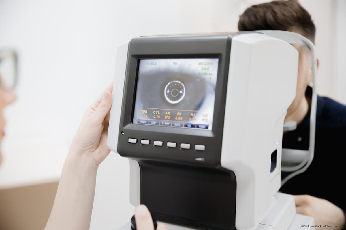
Uveitis is a notoriously challenging disease to manage. Among the leading causes of vision loss worldwide, it can stem from a wide variety of infectious and noninfectious etiologies. Therefore, a multipronged assessment designed to gather as much information as possible regarding the cause and severity of the disease is critical to determine the most appropriate treatment regimen.
Go beyond the referral
In our clinic, an evaluation always begins with obtaining a thorough patient history. While notes from the referring physician are important, they cannot be relied upon solely. We obtain a detailed ocular history, including symptom recurrence, duration, and severity as well as other ocular surgeries or trauma. We review the systemic history, including other illnesses or diseases as well as exposure to uveitis risk factors. Such review is critical, as diseases afflicting other organs in the body such as the kidney or lungs might preclude the use of certain types of medication. Finally, we document the patient’s treatment history, including the dosage of medication(s) and duration of therapy previously prescribed, as well as whether a certain course of therapy failed previously, or the patient experienced recurrences of disease.
Seeing "the big picture"
A thorough clinical examination must include both physical evaluations (ie, slit lamp, indirect ophthalmoscopy, and direct contact lens biomicroscopy) and retinal imaging, considering information garnered from both the anterior and posterior segments. Performing an anterior segment OCT may be helpful for evaluating the cornea. For the posterior segment, ultra-widefield imaging (UWF) is the most efficient and effective tool. While a typical fundus photograph captures 30 degrees of the retina and ETDRS 7 standard fields requires the compilation of 7 of these images to reach approximately 75 degrees, optomap UWF Imaging (Optos plc) captures 200 degrees (82%) in a single capture. No other imaging platform offers the ability to see this far into the periphery. The ability to do so is essential, as lesions often occur in the peripheral retina and their presence would impact treatment decisions.
Figure 1A. Wide-field fundus photographs (A, B) of a 12-year-old Vietnamese American boy with occlusive retinal vasculitis associated with Adamantiades-Behcet Disease showing pale optic nerve, sclerosis of superior nasal retinal arteries, vascular sheathing of temporal inferior arteries, and collateral vessels in the peripheral retina.
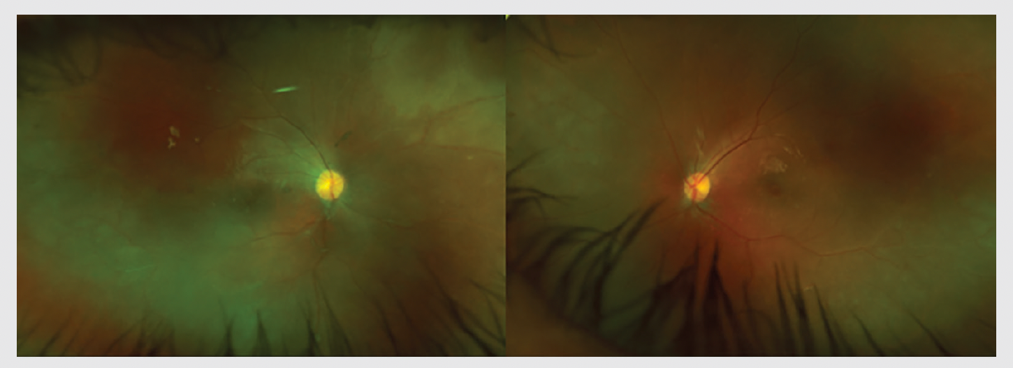
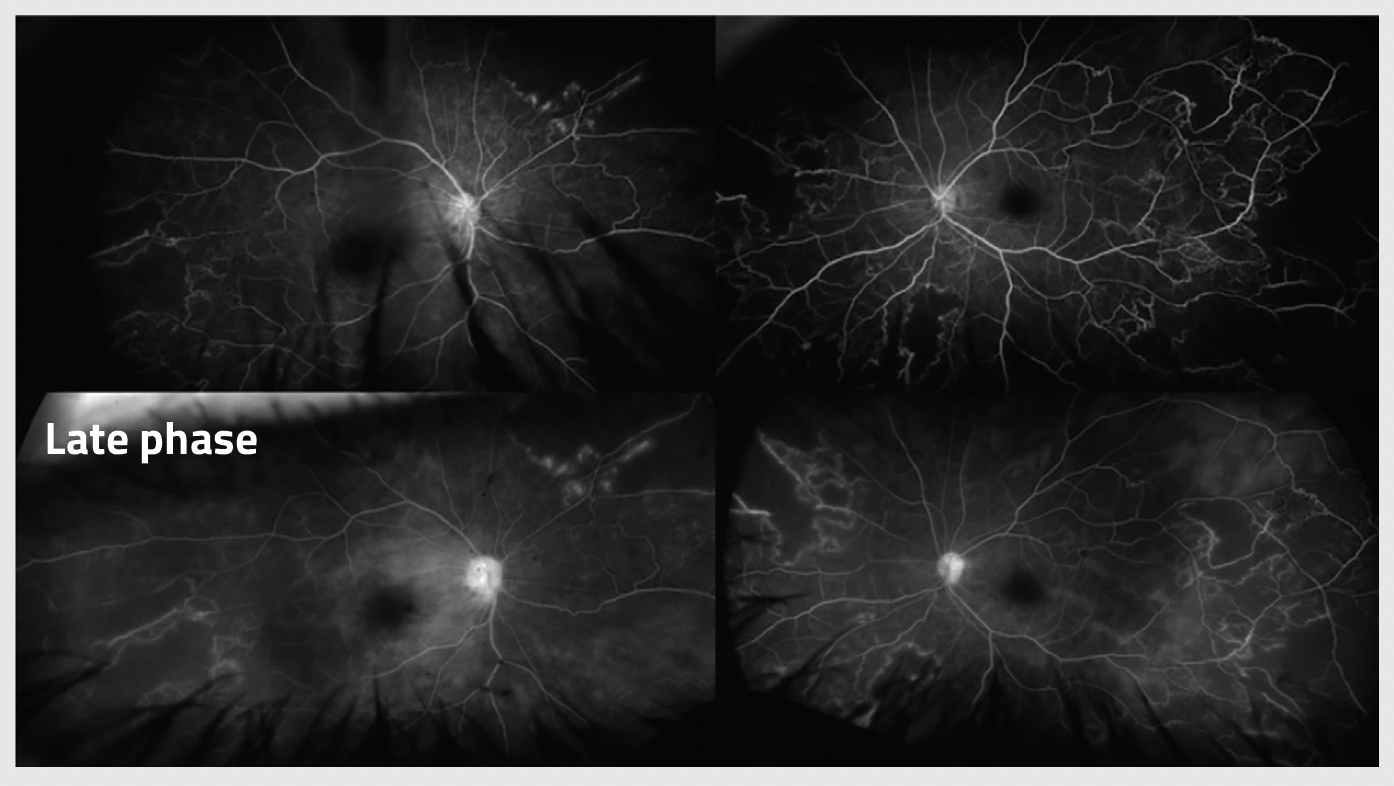
Figure 1B. Corresponding early and late wide-angle fluorescein angiography of the right and left eyes showing optic disc and peri-vascular leakage and widespread retinal ischemia.
Utilization of wide angle imaging was demonstrated for the first time almost 10 years ago in 2 separate studies evaluating the use of UWF technology in the management of noninfectious posterior uveitis and noninfectious retinal vasculitis. The first study, a prospective, observational case series, set out to determine how often disease management decisions changed based on the additional information garnered from UWF fundus images and UWF fluorescein angiography (FA). The results showed that investigator review of UWF and UWF FA led to management changes in nearly half (48%) of the patients evaluated.1 In the second study, evaluation of UWF pseudocolor images led to a 14% increase in management changes, while UWF FA images led to a 51% increase.2
When evaluating a UWF FA image, it is important to look at the perfusion or leakage of the vessels. The presence of retinal vascular leakage and peripheral ischemia can be important signs of significant posterior segment inflammation. These are examples of peripheral findings that would go undetected with traditional fundus photography, but that UWF is able to capture.
UWF retinal imaging also supports disease documentation. Depending on the extent of peripheral pathology identified in an initial capture, it might be prudent to reexamine after a period of time and compare the images to assess disease activity and progression before initiating treatment. Image comparison is especially beneficial when evaluating for both efficacy and safety following treatment initiation.
Understanding potential systemic disease factors
A third critical element of uveitis evaluation is laboratory evaluations. Identifying any potential problems such as low platelet or low white blood cell count, for example, is important when considering the potential root cause of the disease and, subsequently, determining the most appropriate treatment regimen. A thorough initial investigation may also help determine the need to obtain specimen(s) to understand more, such as in the case of intraocular lymphoma, where one needs to look at the vitreous of the eye and not just the fundus.
Conclusion
By improving our understanding of the disease process, we may be better informed regarding our treatment and management decisions, which will hopefully lead to the best possible outcomes for our patients. This approach is particularly important in uveitis, which has a broad array of etiologies, each requiring different therapeutic approaches. In our clinics, this multifaceted approach with the addition of UWF imaging has greatly improved our ability to diagnose and manage patients with uveitis. This critical tool not only helps guide our treatment decisions, but also allows patients to be better informed should the need for intervention or procedures arise.
Quan Dong Nguyen, MD, MSc, FARVO, FASRS
E: ndquan@stanford.edu
Dr. Nguyen serves on the scienti c advisory boards for Affibody, Bausch + Lomb, Regeneron, and Santen, among others. Dr. Ghoraba and Dr. Or have no relevant disclosures.
References
1. Campbell, JP, Leder, HA, Sepah YJ. Widefield retinal imaging in the management of noninfectious posterior uveitis. Am J Ophthalmol. 2012;154(5):908-911.e2. doi:10.1016/j.ajo.2012.05.019
2. Leder HA, Campbell JP, Sepah YJ, et al. Ultrawide- field retinal imaging in the management of non-infectious retinal vasculitis. J Ophthalmic Inflamm Infect. 2013;3(1):30. doi:10.1186/1869-5760-3-30
Newsletter
Keep your retina practice on the forefront—subscribe for expert analysis and emerging trends in retinal disease management.